A Diagram Of Joints And Bones In The Human Body - The 3 Types Of Joints In The Body. At a symphysis, the bones are joined by fibrocartilage. The following activities will help your students learn how the bones, muscles, and joints work together, as well as how to prevent injuries from occurring. Human muscle diagram muscular system muscles of the human body. Altogether, the skeleton makes up about 20 percent of a person's body weight. Male arm and chest muscles labeled chart on white stock.
There are five main shapes of bones: Each hip bone contains three bones— the ilium, ischium, and pubis — that fuse together as we grow older.the sacrum, five fused. The shoulder is comprised of a ball (humerus) and socket (scapula), bones, ligaments, tendons and muscles that move the arms and connect them to the torso. It bears our body's weight and the force of the strong muscles of the hip and leg. The long bones of the body contain many distinct regions due to the way in which they develop.
What Are Bones Bones Of The Human Skeleton Teaching Wiki from images.twinkl.co.uk They provide a great deal of strength to modulate powerful forces between the upper and lower body. This diagram shows the six classes of movable joints in the human body. In the diagrams below, i'll be showing muscle groups in color, with a black line to show the forms that would show through the skin (i also stand in front of a mirror and find each of the muscles shown here in your own body. 612 x 792 jpeg 90 кб. An example of a pivot joint is the joint between the first two vertebrae in the spine. Human muscular system diagram labeled human body muscles. A diagram of joints and bones in the human body / images 04. There are five main shapes of bones:
The following questions are written in language appropriate for sharing with
(benavides 2015) figure 1.5 bones and joints of a human palm and wrist (nanayakkara et al. It bears our body's weight and the force of the strong muscles of the hip and leg. According to the type of tissue at the joint: Cartilaginous joints are where the adjacent bones are joined by cartilage. Yet the hip joint is also one of our most flexible joints and allows a greater range of motion than all other joints in the body except for the shoulder. 612 x 792 jpeg 90 кб. Joints hold the skeleton together and support movement. In humans and other vertebrates, the bones form a framework called the skeletal system that provides structure and shape. There are five main shapes of bones: This comic art reference shows the muscles in the human arm color coordinated. Joints in the human skeleton can be grouped by function (range of motion) and by. Male arm and chest muscles labeled chart on white stock. There are two ways to categorize joints.
Diagrams of human muscles lower arm muscles diagram human muscle. At a symphysis, the bones are joined by fibrocartilage. The skeleton of the human body is made out of bones and the cartilage supporting those bones. The epiphyseal plate of growing long bones and the first sternocostal joint that unites the first rib to the sternum are examples of synchondroses. According to the type of tissue at the joint:
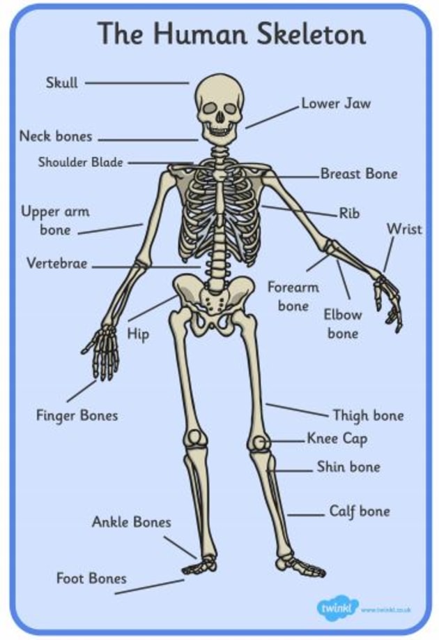
Quickstudy Joints Ligaments Laminated Poster Anatomy Bones Joints Anatomy Human Joints from i.pinimg.com There also are bands of fibrous connective tissue—the ligaments and the tendons—in intimate relationship with the parts of the skeleton. Human muscular system diagram labeled human body muscles. Muscle tendons in the knee joint and the shoulder joint are. The patella and the pisiform bone of the carpals are the only sesamoid bones that are counted as part of the 206 bones of the body. Upper arm diagram filehuman arm bones diagramhebsvg wikipedia. The bones of the leg are the femur, tibia, fibula and patella.the foot bones shown in this diagram are the talus, navicular, cuneiform, cuboid, metatarsals and calcaneus. This diagram shows the six classes of movable joints in the human body. Long (such as the upper arm), short (such as the hand), flat (such as the ribs), irregular (such as the vertebrae) and sesamoid (such as the kneecap).
A joint is an area where 2 or more bones are in contact with each other.
A joint is an area where 2 or more bones are in contact with each other. The shoulder is comprised of a ball (humerus) and socket (scapula), bones, ligaments, tendons and muscles that move the arms and connect them to the torso. The skeleton of the human body is made out of bones and the cartilage supporting those bones. Diagrams of human muscles lower arm muscles diagram human muscle. Long (such as the upper arm), short (such as the hand), flat (such as the ribs), irregular (such as the vertebrae) and sesamoid (such as the kneecap). Joints in the human skeleton can be grouped by function (range of motion) and by. This is a table of skeletal muscles of the human anatomy. They provide a great deal of strength to modulate powerful forces between the upper and lower body. An example of a pivot joint is the joint between the first two vertebrae in the spine. The following activities will help your students learn how the bones, muscles, and joints work together, as well as how to prevent injuries from occurring. The second component of the musculoskeletal system are the joints. Diagram, muscle charts of the human body pt direct, muscular system muscles of the human body, muscles of the torso diagram human anatomy body, printable diagrams of muscles printable diagram, full body muscle diagram tenderness co. Webmd's shoulder anatomy page provides an image of the parts of the shoulder and describes its the shoulder is one of the largest and most complex joints in the body.
This comic art reference shows the muscles in the human arm color coordinated. Yet the hip joint is also one of our most flexible joints and allows a greater range of motion than all other joints in the body except for the shoulder. Elbow joint diagram human elbow elbow joint elbow bones the diagram depicts bones and parts of a human elbow including humerus, trochiea, ulna, radius, head, neck, radial fossa and others. Muscles in human function by producing motion and force and are primarily responsible for: Locomotion is the ability to move from one place to another.
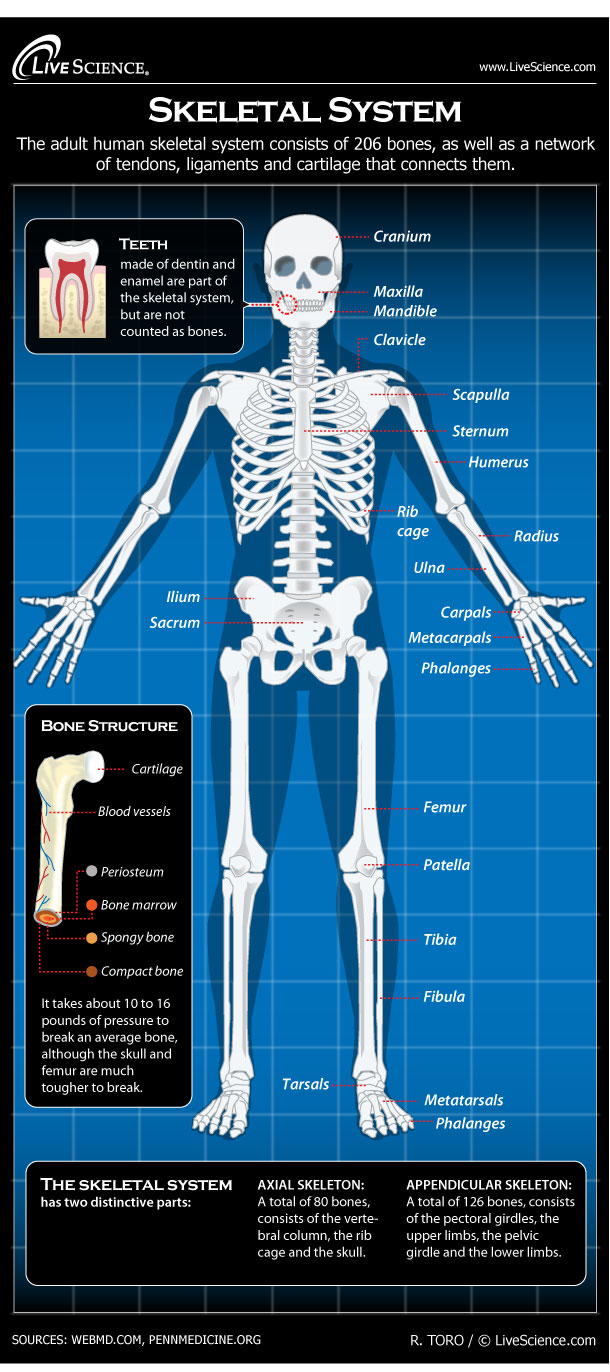
Diagram Of The Human Skeletal System Infographic Live Science from cdn.mos.cms.futurecdn.net Diagram, muscle charts of the human body pt direct, muscular system muscles of the human body, muscles of the torso diagram human anatomy body, printable diagrams of muscles printable diagram, full body muscle diagram tenderness co. It bears our body's weight and the force of the strong muscles of the hip and leg. Skeletal muscles are held to the bones with the help of tendons. Human muscle system diagram human muscular system diagram anatomy | gethumananatomy. The sacroiliac (si) joints connect the sacrum at the base of the spine with the hip bone. Altogether, the skeleton makes up about 20 percent of a person's body weight. This is a table of skeletal muscles of the human anatomy. In human anatomy, the muscles of the hip joint are those muscles that cause movement in the hip.
They provide a great deal of strength to modulate powerful forces between the upper and lower body.
Muscle tendons in the knee joint and the shoulder joint are. It allows us to walk, run, and jump. Upper arm diagram filehuman arm bones diagramhebsvg wikipedia. Joints hold the skeleton together and support movement. In human anatomy, the muscles of the hip joint are those muscles that cause movement in the hip. Cartilaginous joints are where the adjacent bones are joined by cartilage. (benavides 2015) figure 1.5 bones and joints of a human palm and wrist (nanayakkara et al. An example of a pivot joint is the joint between the first two vertebrae in the spine. A joint is an area where 2 or more bones are in contact with each other. The patella and the pisiform bone of the carpals are the only sesamoid bones that are counted as part of the 206 bones of the body. Other sesamoid bones can form in the joints of the hands and feet, but are not present in all people. Male arm and chest muscles labeled chart on white stock. Full body muscular diagram pdf.
About the Author
deary
Author & Editor
Has laoreet percipitur ad. Vide interesset in mei, no his legimus verterem. Et nostrum imperdiet appellantur usu, mnesarchum referrentur id vim.


0 komentar:
Posting Komentar