Anatomy Label Major Arteries And Veins / Major Blood Vessels For Central Line Insertion Stock Illustration Illustration Of Science Medical 41539225. 612 x 513 jpeg 64 кб. The major deep veins of the arm are the radial and ulnar veins, which run along the length of their respective bones and merge at the elbow to form the. Electrical properties of the heart. Match the arteries in column a with the regions supplied in column b. Together, veins, arteries and nerves define neurovasculature.
15.1 abdominal aorta and major branches anterior view. Brachial, radial, and ulnar veins: Roots, trunks, divisions, cords, branches. Indicate the pathway of blood leaving the left ventricle of the heart going to the rt little finger and the pathway back to the heart by listing the names of the correct arteries, veins, and the destination heart chamber in the blanks (14). 612 x 513 jpeg 64 кб.
Cardiovascular System Anatomy And Physiology Study Guide For Nurses from nurseslabs.com Neither the pulmonary artery or vein are listed because they are not systemic; 529 x 644 png 236 кб. 612 x 513 jpeg 64 кб. Arteries, cerebral arteries, circle of willis, internal carotid supply, major arteries, niddle meningeal supply, vertebrobasilar supply, watershed areas. There are three major types of blood vessels: 5 detailed anatomy subclavian origin left from aorta right branching point of brachiocephalic termination of both sides outer border of the first rib axillary outer boarder of first rib termination teres major (both sides) brachial teres major cubital fossa. There are three major branches of the aortic arch: The brachiocephalic artery, the left common carotid artery, and the left subclavian (literally under the clavicle) artery.
Figure 47.14 label the major systemic arteries.
These veins provide superficial venous return. Thoracic aorta, abdominal aorta, iliac arteries veins: Human anatomy for muscle, reproductive, and skeleton. Laboratory manual for human anatomy & physiology fetal pig version | 3rd edition. The external carotid artery supplies the areas of the head and neck external to the cranium. Roots, trunks, divisions, cords, branches. General anatomy and musculoskeletal system. There are about half a dozen arteries to learn. 15.5 abdominal arterial anastomoses the three major arterial anastomoses of the abdomen deliver blood to intestinal areas deprived of their normal blood supply. Place the letter of your choice in the figure 46.11 label the major arteries and veins of the systemic and pulmonary circuits. Illustration depicting main leg arteries (anterior view). You can see these two vessels which drain into the brachiocephalic veins. Lateral pectoral nerves goes through pectoralis major while medial p.n.
There are three major types of blood vessels: There are three major branches of the aortic arch: Learn the major arterial branches off the aorta in the chest, abdomen, and pelvis. And is intended to assist and challenge students in the most effective way to use this workbook is to read reviewing the sectional anatomy and concepts presented the chapters in the. Label the major arteries and veins indicated in.
Major Systemic Arteries Pt 1 Quiz from www.purposegames.com Label the major arteries and veins indicated in. Together, veins, arteries and nerves define neurovasculature. I'm unsure if you're asking about general direction of flow or about memorizing specific names of major arteries and veins. Describe the waveforms and pressures that are seen in each anatomical location during insertion of a pulmonary artery catheter. Veins, blood vessels which return blood to the heart, are different in structure and function from the arteries, which carry blood to the circulation. Anatomy of excitatory and conductive elements: Indicate the pathway of blood leaving the left ventricle of the heart going to the rt little finger and the pathway back to the heart by listing the names of the correct arteries, veins, and the destination heart chamber in the blanks (14). Learn anatomy faster and remember everything you learn.
Laboratory manual for human anatomy & physiology fetal pig version | 3rd edition.
I'm unsure if you're asking about general direction of flow or about memorizing specific names of major arteries and veins. Place the letter of your choice in the figure 46.11 label the major arteries and veins of the systemic and pulmonary circuits. Anatomy of excitatory and conductive elements: Describe the waveforms and pressures that are seen in each anatomical location during insertion of a pulmonary artery catheter. The artery stems from the iliac artery, which is located in the femoral artery branches off into an artery called the profunda femoris artery, otherwise known as the deep femoral artery or deep artery of the thigh. Superior vena cava, azygos, hemiazygos, iliac veins, inferior vena cava nerves: 6 vein names and their branches off the. They accompany the arteries of the. Brachial, radial, and ulnar veins: You can see these two vessels which drain into the brachiocephalic veins. Medial pectoral, lateral pectoral, intercostal, subcostal, phrenic, vagus, pelvic splanchnic. Related posts of anatomy veins arteries diagram. There are about half a dozen arteries to learn.
Place the letter of your choice in the figure 46.11 label the major arteries and veins of the systemic and pulmonary circuits. Illustration depicting main leg arteries (anterior view). These veins provide superficial venous return. They accompany the arteries of the. Roots, trunks, divisions, cords, branches.
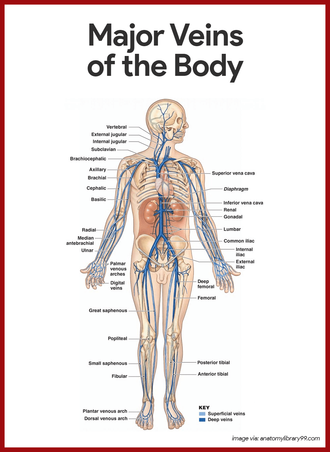
Pulmonary Trunk Radiology Reference Article Radiopaedia Org from prod-images-static.radiopaedia.org Tributaries of the coronary sinus and the anterior cardiac. Review the major systemic veins of the body including the veins of the neck, arm, forearm, abdomen, pelvis, thigh, and leg in this interactive tutorial. Label the major arteries and veins indicated in. Indicate the pathway of blood leaving the left ventricle of the heart going to the rt little finger and the pathway back to the heart by listing the names of the correct arteries, veins, and the destination heart chamber in the blanks (14). The external carotid artery supplies the areas of the head and neck external to the cranium. Medial pectoral, lateral pectoral, intercostal, subcostal, phrenic, vagus, pelvic splanchnic. Brachial, radial, and ulnar veins: 15.1 abdominal aorta and major branches anterior view.
Anatomy of excitatory and conductive elements:
Illustration depicting main leg arteries (anterior view). Major systemic arteries major systemic veins note: 15.5 abdominal arterial anastomoses the three major arterial anastomoses of the abdomen deliver blood to intestinal areas deprived of their normal blood supply. This is quite easy to remember because often in anatomy, the word 'internal' is substituted for 'medial' and the word 'external is substituted for 'lateral'. And posterior view of the heart, arteries, and veins. Systemic arteries and the arterial pathway of blood to. The brachiocephalic artery, the left common carotid artery, and the left subclavian (literally under the clavicle) artery. Roots, trunks, divisions, cords, branches. Table 20.4 defines the major arteries and veins of the pulmonary circuit discussed in the text. Brachial, radial, and ulnar veins: All chapters in this workbook edition of sectional anatomy for imaging professionals correspond with those from the text. Label the major arteries and veins indicated in. 529 x 644 png 236 кб.
About the Author
deary
Author & Editor
Has laoreet percipitur ad. Vide interesset in mei, no his legimus verterem. Et nostrum imperdiet appellantur usu, mnesarchum referrentur id vim.


0 komentar:
Posting Komentar